




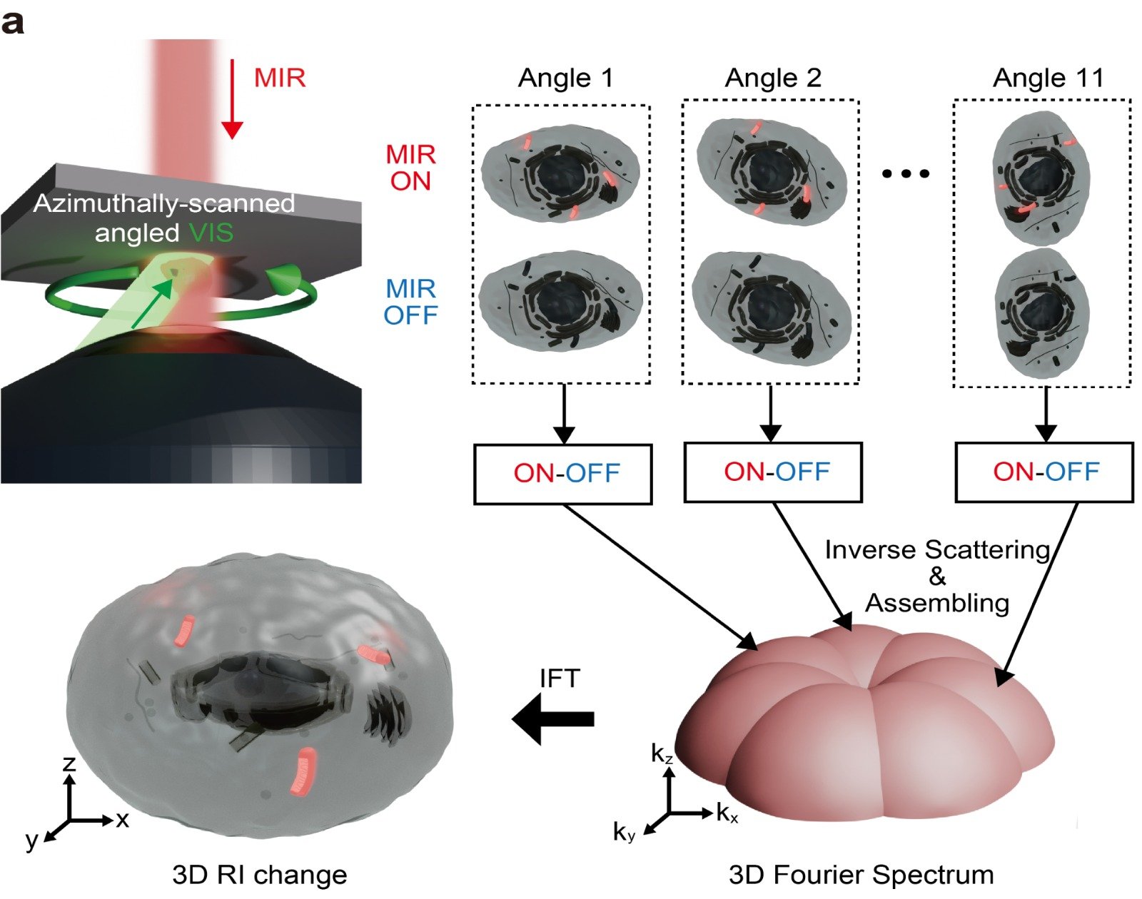
EduAsiaNews, Tokyo – Researchers at the University of Tokyo have developed a high-speed microscopy technique that captures images so rapidly that it can produce “videos” of dynamic processes inside living cells. The team illuminated samples with mid-infrared light to generate photothermal signals, then captured snapshots of temperature changes within cells caused by different molecules. Previous efforts managed to capture just 0.05 frames per second, but the new technique achieves 19.2 frames per second. By capturing images from multiple angles, the researchers can reconstruct 3D “videos” showing ongoing biological processes.
There are many ways to look inside cells to understand how they function, adapt to changes, and respond to treatments. One such technique is label-free mid-infrared photothermal microscopy. It uses mid-infrared light to excite chemical bonds in molecules, each of which responds at a different frequency. These differences can be used to map the internal chemical composition of a cell, which can then be visualized using visible light.
One advantage of this technique is that, unlike some other methods, it does not require dyes that could be toxic to living samples. Combining images captured from different angles also makes it possible to create 3D images. However, capturing images at subsecond intervals has remained a challenge.
Graphic mapping the movement of chemical components inside a cell in 3D.
Tracking movement in 3D space. The graphic shows (a) the three-dimensional movement of lipid droplets over 3.6 seconds and (b) their movement characteristics and how these vary depending on their location within the cell. © M. Fukushima, K. Toda, Y. Sugawara et al. 2026/ Nature Communications, CC BY
“Previously, three-dimensional mid-infrared photothermal tomography was limited to fewer than 0.05 frames per second, mainly because signal averaging and sequential measurements at different illumination angles required considerable time,” explained Masato Fukushima, the study’s first author and a doctoral student in the Department of Physics. “However, we have developed a new technique that achieves 19.2 frames per second, allowing us to obtain a complete three-dimensional image every 52 milliseconds, approximately 400 times faster than previous approaches.”
The team overcame the speed limitation by using a spatial light modulator — a device that precisely controls light properties, such as the phase of its waves, to direct light in different directions — to change illumination angles rapidly. They also maximized the signal-to-noise ratio for each image to ensure the clearest possible results. The resulting system is called video-rate mid-infrared photothermal optical diffraction tomography (MIP-ODT).
A series of images showing the chemical distribution inside a cell at different depths.
Photothermal images. The images show differences in mid-infrared light absorption depending on intracellular structures. © M. Fukushima, K. Toda, Y. Sugawara et al. 2026/ Nature Communications, CC BY
Using MIP-ODT, the researchers were able to observe lipid droplets inside cells, which play a role in energy regulation and lipid storage, and track their movement in three-dimensional space. This revealed transport behavior that is difficult to detect through two-dimensional imaging alone.
“Our research transforms this technique from a method primarily used to observe relatively static samples into a tool capable of quantitatively investigating rapid biological processes in three dimensions,” Fukushima said.
The team anticipates several promising future applications for the technology. One potential use is the study of biomolecular condensates, membrane-free structures inside cells that contain proteins and nucleic acids. Abnormal behavior in these condensates has been linked to neurodegenerative disorders, so investigating how they grow and change could help researchers develop new treatments.
“The next step is to address fundamental biological questions that conventional techniques cannot explore. The main strength of label-free chemical imaging is its ability to provide quantitative information about the chemical composition of living cells,” said the study’s senior author, Professor Takuro Ideguchi of the Graduate School of Frontier Sciences.
“By visualizing cellular dynamics in three dimensions and over time, we aim to advance toward a quantitative, measurement-based understanding of life,” he added.